

Microscope Monday 06-18-2024
Microscope Monday 06-18-2024
A special stain known as Warthin-Starry

The world of surgical pathology is more than just looking at pink and purple in the microscope. There is also a vast array of special stains that allow us to identify certain characteristics of the tissue, often involving microorganisms. The Warthin-Starry stain is one that stains most of the tissue a bright yellow-orange-gold kind of color whereas the nuclei of cells and microorganisms usually stain very dark brown to black. I ordered this WS on a lymph node I was looking at because I wanted to rule out a disease like Cat Scratch Disease:
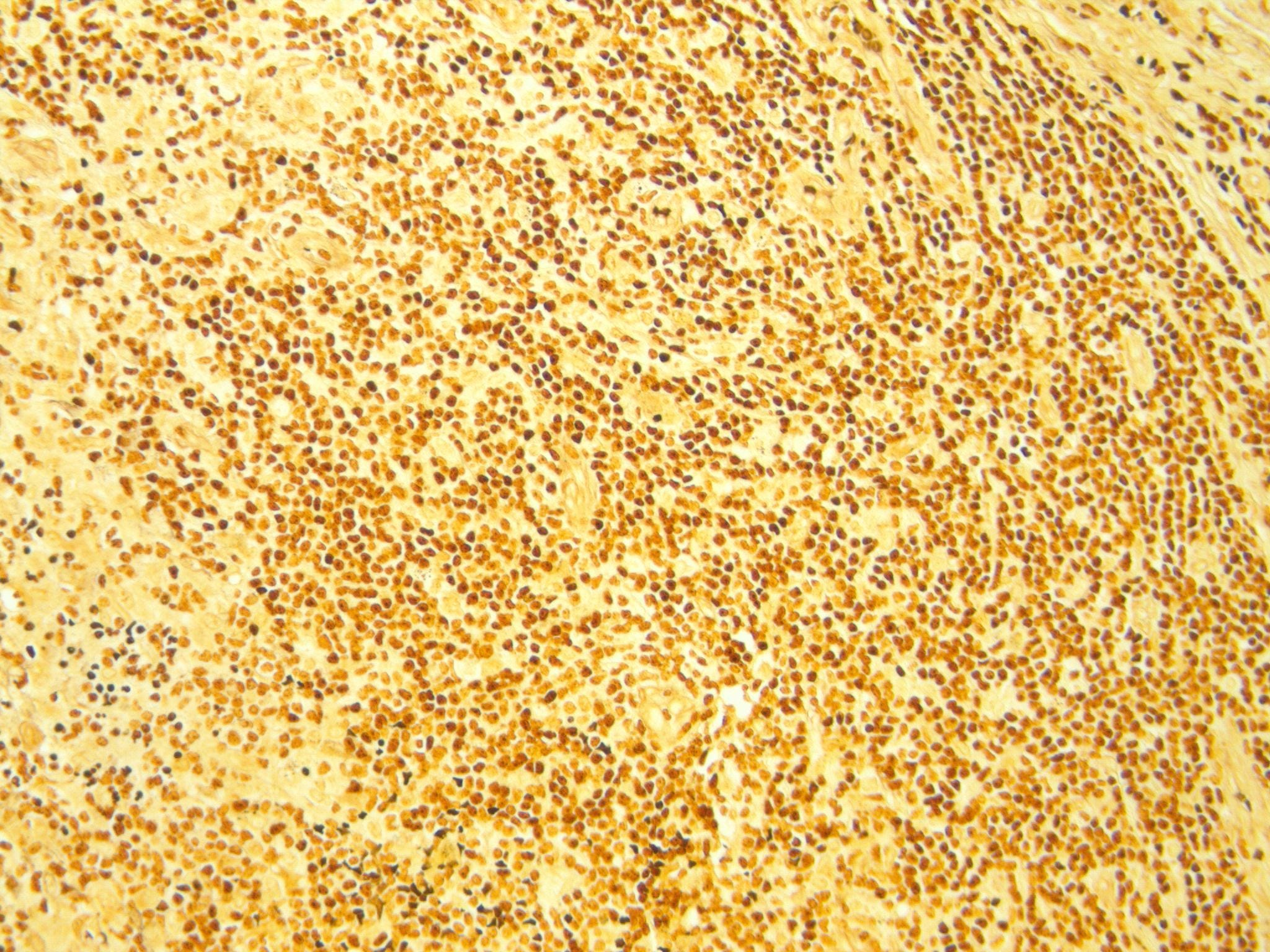
This stain is negative for organisms, and it ended up just being a a lymph node with inflammation. One of my favorite uses for the stain was to look for Helicobacter pylori bacteria in gastric glands. This infection is a very treatable cause of gastritis, and curing an H. Pylori infection will not only make the patient feel a lot better, but it will also decrease their risk of gastric lymphoma and gastric carcinoma. Here is what a positive stain looks like in the stomach glands:
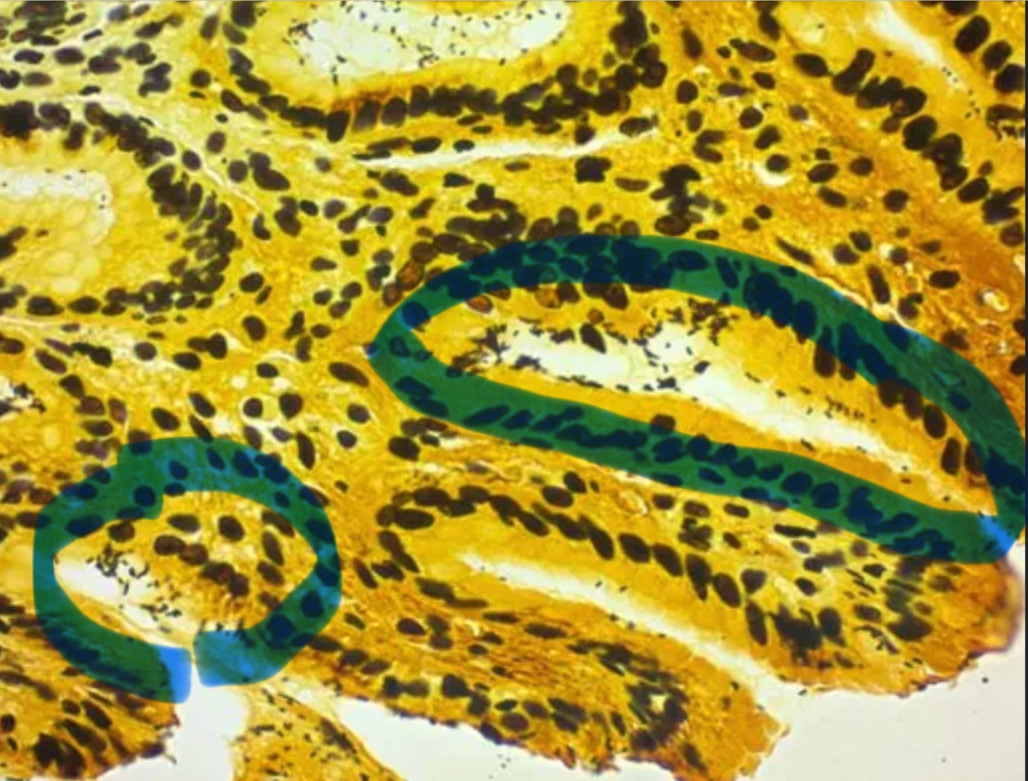
Those little black specks are the organisms. A this microscopic field is around 200x, so you can see they are quite small. It should be noted that there are other stains to look for these organisms, including Giemsa and H. Pylori immunohistochemical stain. The latter is by far the most common used these days, but I still prefer Warthin-Starry for this purpose.
This is what can be fun about pathology: making the decision to look a little deeper at a tissue, order a stain, and be able to result in a patient cure.
—DLW
I’m having lots of blood tests these days. These posts are really helpful for me to understand what goes on with my testing. Thanks for sharing your knowledge.
Learning about the Warthin-Starry stain features, seeing the slide indicating the H.
pyloric bacteria invading the gastric glands, was mesmerizing! 👏💜🤗
I really enjoy studying your pathology site.
Thank you!