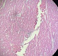
Observe the following image and think about the different structures:
See anything interesting? If you want to quiz yourself, ask yourself what tissue or organ this is from. Answer coming after next image. Let me show you what I think is most interesting here:
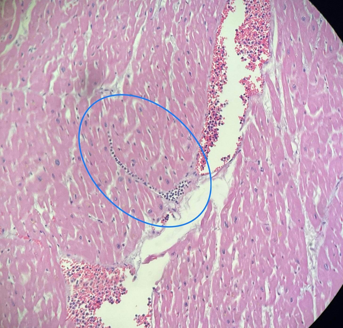
Inside that blue oval is a tributary of a vein (this is a capillary, but seeing it arise directly from the larger vein made me want to use the term tributary…which is not medical use) arising from a larger vein. It’s in a longitudinal section and to see this length of a blood vessel in a microscopic section is very unusual. What’s more is it is filled with white blood cells lined up cell by cell, another unusual finding. To give you a sense of scale, each one of those white blood cells is about 10 - 15 microns, and 1 micron is 1/100th of a millimeter. So that blood vessel is at most 15 microns in diameter.
By the way, this is heart muscle, aka myocardium. The presence of these white blood cells (they’re neutrophils, actually, a type of cell that is active during acute situations) does not indicate infection in this instance; rather it is due to the extreme physiological stress at the time just before dying, and the body is releasing lots of epinephrine and norepinephrine (common use = adrenaline, although I do not use that term) and those chemicals actually cause an increase in white blood cell count acutely. So, it is common to see this sort of thing in autopsy microscopy.
Hope you enjoyed this little exposition.


That's actually cute. A little wee venule in complete cross section.